
The progressive reduction of MAIT cells is associated with disease severity in liver failure patients
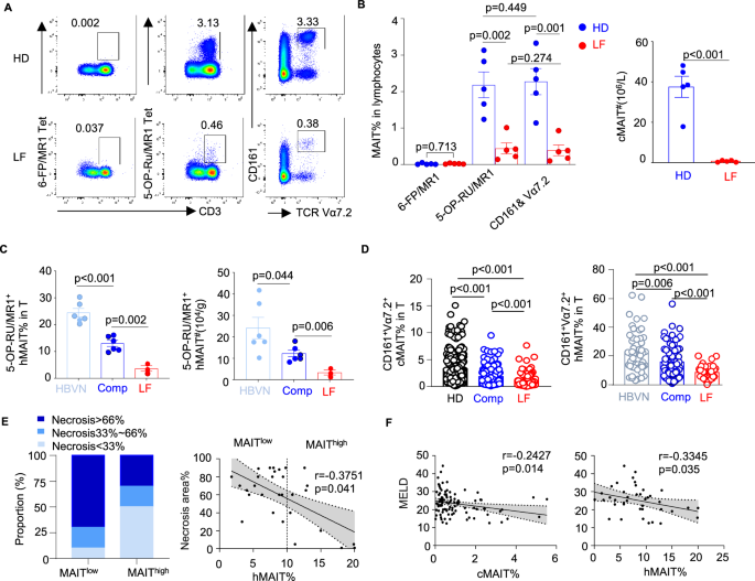
A cohort study was conducted to unveil MAIT cell profiling in chronic HBV-infected patients with liver failure (LF) undergoing artificial liver support or liver transplantation. Healthy donors (HD), HBV-negative carcinoid patients (HBVN), and HBV-positive patients with compensated liver function (Comp) served as controls (Supplementary Fig. 1A). MAIT cells were identified through 5-OP-RU/MR1 tetramer staining or co-staining with antibodies against CD161 and TCRVα7.2 (anti-CD161/Vα7.2) (Fig. 1A and Supplementary Fig. 1B–D). 5-OP-RU/MR1 tetramer-positive T cells and anti-CD161/Vα7.2 co-stained T (CD161+Vα7.2+ MAIT) cells exhibited consistent decreases in the blood and liver of LF patients compared to HD, HBVN, and Comp groups (Fig. 1B–D), showing a predominant CD8-positive population (Supplementary Fig. 1B).
A, B Representative plots (A) and summarized graph (B, left panel) for MAIT cell staining by 6-FP/MR1 tetramer, 5-OP-RU/MR1 tetramer, and/or anti-CD161/TCRVα7.2 antibodies from healthy donors (HD, n = 5) and liver failure patients (LF, n = 5). Summarized graphs (B, right panel) of the numbers of circulating MAIT cells (cMAIT) from HD (n = 5) and LF (n = 5). Statistical significance was assessed by the two-sided student’s t test. C Summarized graphs of frequencies and numbers of hepatic MAIT cells (hMAIT) staining by 5-OP-RU/MR1 tetramer from HBV negative carcinoid patients (HBVN, n = 6), chronic hepatitis B patients with compensated liver function (Comp, n = 6), and liver failure patients (LF, n = 3). Statistical significance was assessed by a two-sided unpaired t test between the two groups. D Summarized frequencies of circulating CD161+TCRVα7.2+ MAIT cells (cMAIT) in T from HD (n = 292), Comp (n = 180) and LF (n = 107) and frequencies of hMAIT from HBVN (n = 50), Comp (n = 70) and LF (n = 42). Statistical significance was assessed by the Kruskal-Wallis test followed by Dunn’s test. E The proportion of liver failure patients with different degree of necrosis in groups with MAIT cell frequencies higher than 10% (MAIThigh, n = 10) and lower than 10% (MAITlow, n = 20) (left panel); Spearman correlation between frequencies of hMAIT and necrosis areas of liver tissue from liver failure patients (n = 30, right panel). F Spearman correlation between the scores of the model for end-stage liver disease (MELD) and ratios of cMAIT (n = 102) and hMAIT (n = 40) from liver failure patients. Data are presented as mean ± SEM. n-values represent biological replicates. Source data are provided as a Source Data file.
Albeit the frequencies of circulating (cMAIT) and hepatic MAIT (hMAIT) cells from the Comp group already reduced as compared to those from HD or HBVN, MAIT cells further decreased and displayed a higher apoptosis rate in liver failure patients (Supplementary Fig. 2A and B). Of note, cMAIT and hMAIT frequencies reduced to a greater extent in liver failure patients presenting with larger liver necrotic areas (Fig. 1E), more frequent cholestasis (Supplementary Fig. 3A and B), higher scores of model for end-stage liver disease (MELD) prioritizing patients for liver transplant (Fig. 1F), higher value of histological activity index (HAI) reflecting tissue necroinflammation (Supplementary Fig. 3C), and/or multiple complications (Supplementary Fig. 3E). In contrast, nucleos

