
Assuming that the level of abdominal fat that has been decided is high, it is assumed that it is associated with a decrease in renal function. In this study, before donation and (analysis of MGFR standardized MGFR, and (analysis of BSA standardized MGFR), between the higher fat regions associated with renal functions with low renal function. , It has been found that there is a significant relevance between the CT determined abdominal fat area and the renal function, to elucidate the effects of the body composition (measurement) for renal function. 。
Before the donation, all abdominal fat parcels showed a significant association with renal function, and the MGFR value was lower due to the high value of the CT -derived abdominal fat area. According to research among healthy people, visceral obesity was independently related to renal dysfunction in all ages and two gender, except for men under the age of 45.18。 VAT, Sat, and IMAT are related to the decrease in renal function and/or chronic kidney disease in general groups.19And the VAT area has an independent influence on the estimated GFR (EGFR) level in a healthy woman's research.20。 In addition, the visceral fat area of the baseline was associated with a healthy protein urine.twenty one。 Interestingly, the donor, which is particularly about the minimum fat index, had a higher MGFR level in this study compared to donors, which had a high level of fat index.
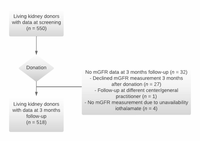
After the donation, three months follow -up, the fault shooting measured abdominal fat area was strongly related to the lower (BSA standardization) MGFR level. Furthermore, don, a higher Don -pillow statue, abdominal fat tissue index levels, have experienced a significant decrease in renal functions between donations and three months after donation. A study of Japanese kidney donors showed that the renal function 12 months after renal kidney surgery, the donor in the VAT area higher than the low VAT area is lower.twenty two。 The measurement of the posterior peritoneal fat tissue was significantly correlated with the decrease in EGFR in the first and six months after donation compared to the EGFR before the donation.twenty three。 Thus, donors, which have a high level of abdominal fat area, seem to start renal functional orbital after kidney due to low renal function. It is interesting to know how the abdominal fat level and the renal function after the DO develop long -term after donation. In this study, 57 % of the research groups were able to use renal functional data five years after donation. The analysis using these long -term renal functional data suggests that the level of the fat region of the kidneys five years after the fault shooting is high, but the further research on the long -term consequences of kidney donation is complete. It suggests that it is essential to this association.
The negative relevance between IMAT and the renal function was an interesting discovery. IMAT is an indicator of flexible pulmonary and skeletal muscles, and is a predictor of a deviated course in many patient groups.twenty four,,twenty five,,2627。 Most studies use the attenuation or muscle density of skeletal muscle radiation measured by HU as a substitute marker for muscular bone disease. The measurement of adipose tissue in the skeletal muscles on CT scan is a new method for evaluating fascia, and may be a promising method in research on fascia. As far as we know, this is the first study that shows the relationship between fascia and renal function in a healthy kidney donor. Further studies, including long -term clinical results, such as the onset of kidney disease, are required to establish muscle internal fat tissue and fascia cut -off value.
The relationship between the abdominal fat area and the renal function before and after donor kidney detection was the most obvious when using the standardized MGFR for the BSA. In the secondary analysis using un standardized MGFR, the abdominal fat area and the relevance of MGFR were added to the regression model. We have added weight to take into account the size of the body. It is known to affect renal function17。 When the BSA GFR is standardized, the effect of the body size is removed, and it is a known methodological practice, but it is currently discussing the great consequences in extreme groups of body sizes.28。 For screening guidelines on dedication of kidney life at our center (for example, in the case of BMI> 30 kg/m, advice to lose weight2 Exclude in the case of BMI≥ 35kg/m2), The research group of this research was composed of individuals of relatively “normal” body size. The standardization of BSA has little effect on the GFR level of an individual with such a “normal” body size, and can more specific the relationship between abdominal fat area and renal function regardless of the whole body. I have sex.29。
Mechanisms that may be under the negative relevance between abdominal fat tissue and renal function include direct and indirect effects. The direct interaction between the fat tissue and the kidneys is called the “fat axis” and plays an important role in maintaining normal renal function. Poglim tissue secretes a large number of factors that act aggressively in endocrine systems.30,31。 The normal level of these fat tissue factors is important in maintaining renal function.31。 Excessive calorie intake causes fatal hypertrophy or hyperplaces, causes a number of processes that cause fat -derived molecules and metabolites for coordinates, causing oxidative stress, (chronic) inflammation, and kidney fibrosis, Crys renal disorders31。
It also has an indirect effect. Increasing the amount of visceral fat tissue is related to the state of metabolic syndrome and diabetes.32,,3334Danger factor for developing CKD35,,3637。 Other fundamental mechanisms may be associated with the mechanical stress of the kidney, for example, by the pressure on the fat tissue around the kidneys. The increase in fat tissues around the peritoneal can cause kidney damage due to real and direct obstruction of blood vessels, which may increase sodium reabsorption and then cause hypertension.38,39。 Furthermore, the actual compression of the kidneys leads to an increase in interstitial static water pressure, causing the kidney blood flow and the progress of kidney disease.37,,3839。
Many are unknown about the relationship between abdominal fat tissue and kidney function, and mechanisms that may be under the basis of future research. Future research may potentially incorporate (pathological) parameters, such as interstitial fibrosis, urinary tube atrophy, and proto -biological peripheral fat tissues. Elucidate these connections.
The strength of this study includes a relatively large cohort size, renal function measurement (in contrast to estimation), and limited data (less than 5 %). The restriction is a limited number of commonization, as the lack of obese donors, most of the participants are European, single institutions, and retrospective designs. The oldest time after the donation when the MGFR was measured was three months after control. This obstacle analysis of the remaining single kidney MGFR of the remaining kidney for the first few months after donation.
This study shows that higher levels of abdominal fat, as measured by CT analysis, are related to the low renal function after Do. The magnitude of the abdominal fat area to the renal function after short -term donation may seem smaller than that of factors such as age and MGFR before donation, but it is possible to optimize body formation. It is one of the few factors. Before donation. This study also raises questions about the use of BMI as a scale of gold standard.3,,45 To screen donors to do not provide information about the abdominal fat area. BMI did not show the relevance to MGFR before donation. CT analysis, which is performed on a daily basis at most centers as part of the screening of donations, may be a more effective way to evaluate health risks related to body composition. It is necessary to translate this knowledge into clinical medical treatment, determine the effectiveness of CT -derived body composition measurement in the Donar cleaning guidelines, and investigate the potential advantages of reduced abdominal fat in raw donors.
This study indicates that the higher abdominal fat area measured by CT analysis is low in renal function after screening and after kidney. The results of this study use it to elucidate the effects of body composition on the renal function of living kidney donors. Further surveys are essential to investigate the relationship between a variety of (living kidney donors) groups, especially in obese, and the relevance of fat tissues measured through renal function. Furthermore, for example, the effects on the radiological evaluation of fat tissue around the kidney may be considered to be more interested in expanding the understanding of living kidney donations and the transformation of the kidneys.
 Arabic
 Chinese (Simplified)
 Dutch
Dutch English
 French
 German
 Italian
 Portuguese
 Russian
 Spanish