
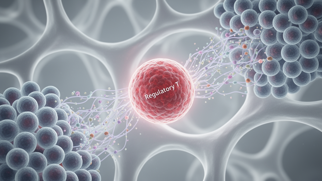
Regulatory T cells actively shape the bone marrow microenvironment, establishing a delicate cellular balance that maintains healthy blood cell production while simultaneously presenting distinct vulnerabilities in malignant and autoimmune conditions. In a comprehensive review published in Frontiers in Cell and Developmental Biology, researchers Jiashuo Cheng, Qian Wang, Zhifeng Gu, and Chen Dong from Nantong University evaluated the intricate molecular and cellular crosstalk connecting regulatory T cells, commonly designated as Tregs, with hematopoietic stem cells, mesenchymal stromal cells, and neighboring immune populations. By synthesizing findings from literature spanning January 2000 through June 2026 across PubMed, Web of Science, and Embase, the authors demonstrated that bone marrow Tregs do not act merely as passive circulating bystanders. Instead, these specialized cells reside in dedicated anatomical compartments where they exert direct biochemical influence over stem cell quiescence, stromal differentiation, and local lymphoid development.
Under normal physiological conditions, regulatory T cells populate the bone marrow niche in substantial proportions, representing approximately 30 to 40 percent of CD4-positive T cells in mouse models. This physical architecture enables regulatory cells to modulate stem cell behavior via contact-dependent surface interactions and locally secreted paracrine factors. Murine niche-associated Tregs expressing high levels of CD150 protect hematopoietic stem cells against oxidative stress by metabolizing extracellular nucleotides. Through the concerted enzymatic activities of surface ectonucleotidases CD39 and CD73, these cells generate extracellular adenosine, dampening harmful reactive oxygen species and preserving stem cell dormancy. In addition, experimental models show that connexin 43 gap junctions facilitate the direct intercellular transfer of cyclic adenosine monophosphate from Tregs to aged hematopoietic stem cells, reducing apoptotic priming and sustaining niche stability.
The bidirectional dialogue between regulatory T cells and mesenchymal stromal cells provides an essential structural pillar supporting normal hematopoiesis. Stromal elements release critical chemoattractants, notably CXCL12, which binds to CXCR4 receptors abundantly expressed on bone marrow Tregs. Genetic deletion experiments in mice reveal that selectively removing CXCR4 in adult Tregs diminishes their presence specifically within the marrow without causing widespread depletion across the spleen or skin. Reciprocally, bone marrow Tregs secrete interleukin-10, an anti-inflammatory cytokine that restrains uncontrolled mesenchymal stromal cell proliferation and guides stromal lineage differentiation while maintaining their capacity to engraft hematopoietic progenitors. Mesenchymal stromal cells likewise preserve Treg identity, releasing extracellular vesicles loaded with regulatory microRNAs, such as miR-181a-5p, which help sustain stable expression of the lineage-defining transcription factor FoxP3 in CD4-positive T cells.
Beyond stem and stromal cell maintenance, bone marrow Tregs enforce strict checkpoints on lymphoid and myeloid differentiation within the hematopoietic niche. In mouse loss-of-function models, the complete absence or acute depletion of Tregs causes residual effector T cells to release excessive inflammatory cytokines, including granulocyte-macrophage colony-stimulating factor, tumor necrosis factor, and interleukin-6. This unchecked inflammatory cascade skews blood production toward granulocyte development at the direct expense of B-cell lymphopoiesis. Furthermore, inducible deletion of CXCR4 in adult mouse Tregs triggers a marked local accumulation of bone marrow B-1 cells, leading to increased synthesis of self-reactive immunoglobulin M and elevated autoantibody levels, whereas peripheral B-1 cell populations remain unaffected. Tregs also maintain close spatial associations with bone marrow plasma cells and CD11c-positive dendritic cells. Depleting Tregs diminishes marrow plasma-cell reservoirs, whereas disrupting Treg-expressed CTLA-4 promotes plasma-cell accumulation, demonstrating that local regulatory networks govern long-term antibody-producing niches.
Epigenetic mechanisms govern the functional stability and specialization of bone marrow regulatory T cells, dictating whether they maintain immune quiescence or succumb to pathological reprogramming. The lineage fidelity of Tregs relies heavily on the demethylation of conserved non-coding sequence 2 within the FoxP3 locus, known as the Treg-specific demethylated region. Incomplete demethylation of this locus compromises FoxP3 expression and impairs suppressive potency under inflammatory pressure. In parallel, histone-modifying enzymes such as enhancer of zeste homolog 2, or EZH2, play decisive roles in enforcing transcriptional repression. Operating as the catalytic engine of polycomb repressive complex 2, EZH2 catalyzes the trimethylation of histone H3 at lysine 27. Upon Treg activation, EZH2 cooperates with FoxP3 to silence conventional effector programs. Genetic ablation of EZH2 destabilizes activated Tregs, precipitating inflammatory phenotypes in preclinical models, which illustrates how local metabolic shifts, hypoxia, and chronic inflammatory signals can induce enduring changes in regulatory cell identity.
In hematological malignancies such as multiple myeloma and acute myeloid leukemia, malignant clones exploit regulatory T cells to construct protective immunosuppressive sanctuaries. Within multiple myeloma, clinical observations show alterations in Treg abundance across monoclonal gammopathy of undetermined significance, smoldering myeloma, and active disease. In mobilized peripheral blood stem cell grafts obtained after induction chemotherapy, elevated frequencies of TIGIT-positive and CD39-positive Tregs correlate with premature disease progression in cytogenetically high-risk patients. Murine myeloma models establish that selective Treg depletion during stem cell mobilization unleashes robust, polyfunctional CD8-positive T-cell responses that significantly improve post-transplant tumor control. Malignant marrow environments also foster the recruitment of specialized conventional type 1 dendritic cells displaying high CD103 and low interleukin-12, which drive the accumulation of both Tregs and exhausted TIM-3-positive, PD-1-positive cytotoxic T cells.
In acute myeloid leukemia, transformed blasts actively recruit, generate, and reprogram bone marrow Tregs through convergent metabolic, receptor-mediated, and vesicular pathways. Leukemic blasts express surface PD-L1 and ICOSL, signaling through PD-1 and ICOS to induce and expand functional CD4-positive CD25-positive FoxP3-positive Tregs. In parallel, leukemia-derived extracellular vesicles bearing 4-1BBL transfer functional signals that enhance Treg suppressive activity and upregulate surface CD39 and CCR8. Furthermore, leukemia-derived interferon-gamma induces indoleamine 2,3-dioxygenase 1 in surrounding mesenchymal stromal cells, driving tryptophan depletion and local kynurenine accumulation that accelerates Treg conversion.
In sharp contrast to hematological malignancies, immune-mediated bone marrow failure syndromes, including aplastic anemia and immune thrombocytopenia, are characterized by severe numeric deficiencies or qualitative defects in regulatory T cells. In aplastic anemia, bone marrow and peripheral blood exhibit reduced proportions of CD4-positive CD25-positive Tregs, and crossover coculture experiments confirm intrinsic functional impairments that prevent adequate suppression of autologous effector T cells. This breakdown allows unrestrained activation of CD8-positive cytotoxic lymphocytes alongside Th1 and Th17 cells, unleashing high levels of interferon-gamma and tumor necrosis factor that destroy early hematopoietic progenitors. Histological evaluations in pediatric aplastic anemia reveal marked reductions in FoxP3-positive cells residing near the endosteal bone surface, coinciding with severe depletion of long-term hematopoietic stem cells. Persistent Treg deficits frequently continue even after clinical immunosuppression, suggesting that enduring regulatory instability may perpetuate chronic marrow damage.
Similarly, patients with active immune thrombocytopenia display significant reductions in bone marrow CD4-positive CD25-positive FoxP3-positive Treg frequencies compared with paired peripheral blood and healthy control marrow. Defective stromal support contributes substantially to this localized deficiency, as bone marrow mesenchymal stromal cells from affected individuals exhibit increased senescence and apoptosis, which cripples their ability to induce functional Tregs in vitro. This cellular defect can be partially counteracted in experimental settings by exposing patient-derived stromal cells to platelet-derived growth factor-BB or mesenchymal stem cell-derived exosomes carrying regulatory microRNAs. Additionally, nonconventional regulatory subsets, such as interleukin-35-producing iTr35 cells, are depleted in the marrow of treatment-naive primary immune thrombocytopenia patients, accompanied by reduced receptor expression on developing megakaryocytes. Because interleukin-35 signaling through AKT promotes megakaryocyte maturation and platelet generation, loss of this regulatory subset directly links immune dysregulation to impaired platelet production.
The contrasting pathological roles of regulatory T cells across hematologic conditions demand opposing therapeutic strategies tailored precisely to disease context, cellular subset, and anatomical compartment. In bone marrow failure, investigational efforts center on adoptive cell therapy, cytokine expansion, and stromal conditioning to reconstitute regulatory coverage. Early clinical trials assessing autologous regulatory T cells expanded with interleukin-2 and rapamycin demonstrated feasibility and yielded partial hematologic responses in three of six refractory aplastic anemia patients, while phase I studies of allogeneic umbilical cord blood-derived products confirmed acceptable safety profiles.
Conversely, breaking immune tolerance in hematologic malignancies requires the selective removal or functional attenuation of tumor-promoting regulatory T cells without triggering catastrophic systemic autoimmunity. Therapeutic candidates include engineered antibody-drug conjugates such as PF-08046032 and afucosylated anti-CD25 antibodies like RG6292, which selectively deplete high-affinity CD25-positive Tregs while enabling natural killer cell-mediated destruction of CD25-bearing leukemic blasts. Disrupting specific checkpoint cascades, including the TIGIT, TIM-3, and BET bromodomain pathways, similarly attenuates Treg viability and restores cytotoxic antitumor vigilance in preclinical models. However, because systemic ablation of FoxP3-positive regulatory cells elicits severe multiorgan autoimmune inflammation in animal models, translational development requires precise timing and niche-restricted targeting to dismantle localized immunosuppressive barriers while safeguarding long-term stem cell survival.
Subject of Research: Bone marrow regulatory T cell biology and hematologic disease remodeling
Article Title: Regulatory T cells in the bone marrow microenvironment: homeostatic crosstalk, disease remodeling, and therapeutic opportunities
Article References: Cheng, J., Wang, Q., Gu, Z., & Dong, C. (2026). Regulatory T cells in the bone marrow microenvironment: homeostatic crosstalk, disease remodeling, and therapeutic opportunities. Frontiers in Cell and Developmental Biology, 14, Article 1990584. https://doi.org/10.3389/fcell.2026.1990584
Image Credits: AI Generated
DOI: 10.3389/fcell.2026.1990584
Keywords: Regulatory T Cells, Bone Marrow Microenvironment, Hematopoietic Stem Cells, Aplastic Anemia, Multiple Myeloma, Acute Myeloid Leukemia, Myelodysplastic Syndromes, Immune Thrombocytopenia, Mesenchymal Stromal Cells, CXCR4, Immunotherapy, CD25, FoxP3, Hematopoiesis, Cellular Biochemistry, Stem Cell Niches, Cytokines, Tumor Microenvironment

